
Central vision loss affects more Americans than you might expect. Up to 90 million adults over age 40 experience vision problems, with age-related macular degeneration representing the most common cause of central vision deterioration. Your visual system commands more than one-third of your brain’s processing power, making central vision essential for reading, recognizing faces, and completing detailed tasks that define daily independence.
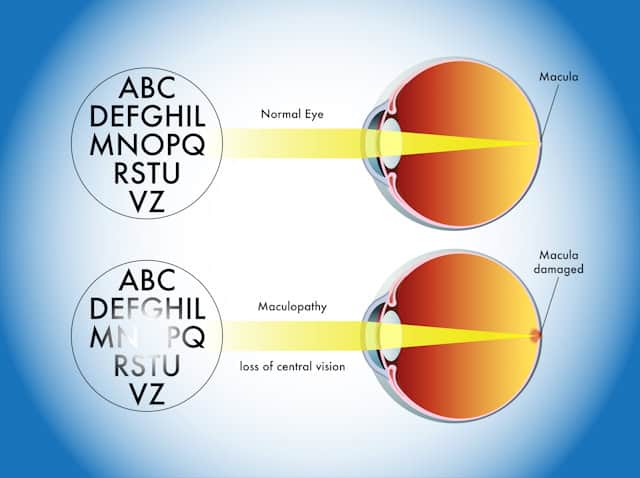
What makes central vision loss particularly concerning is how it strikes at the heart of activities you rely on most. When the macula, the crucial central area of your retina, becomes damaged, simple tasks like reading a prescription bottle or recognizing a loved one’s face become challenging or impossible.
The good news: understanding central vision loss and accessing proven treatments can help you maintain your independence and quality of life.
Early detection can preserve the vision you have while preventing further deterioration.
This guide reveals the conditions that threaten your central vision, explains how doctors diagnose these sight-stealing diseases, and outlines the treatment options available when vision changes occur. You’ll discover practical strategies for adapting your daily activities and learn how early detection can preserve the vision you have while preventing further deterioration.
Whether you’re experiencing the first signs of central vision problems or supporting someone who is, this information can make the difference between losing independence and maintaining an active, fulfilling life.
What Is Central Vision Loss?
How central vision works
Your central vision operates through a remarkable section of your eye called the macula, a small but powerful area at the center of your retina. Think of your retina as a movie screen where light creates the images you see. When light enters through your pupil, your lens focuses it onto this thin layer of light-sensitive cells at the back of your eye.
The macula handles the center of your visual field, enabling you to see color and fine details with precision. This specialized area, measuring only about 6 millimeters across, processes the sharp, detailed images essential for reading, driving, and recognizing faces. Without a functioning macula, activities requiring fine visual discrimination become difficult or impossible.
Your retina converts light into electrical signals that your optic nerve transmits to your brain for processing. While surrounding retinal tissue maintains your peripheral vision, the macula specifically manages what you see when looking straight ahead. This concentration of visual processing power explains why macular damage creates such devastating effects on daily functioning while often leaving side vision intact.
Types of central vision loss
Central vision loss manifests as one or more dark or blurry spots in your field of vision, which may expand or multiply over time. You might notice a dark spot, smudge, or blurred area directly in the center of your visual field. Eye doctors call these gaps scotomas, with central scotomas specifically affecting the macular visual field around your fixation point.
The progression varies dramatically between individuals. Some experience sudden onset while others develop gradual deterioration. Central scotomas create major visual acuity loss, making everyday tasks like reading extraordinarily difficult. When a central scotoma covers your reading visual field, traditional reading becomes impossible without assistance.
However, your visual system possesses remarkable adaptability. You can learn eccentric fixation, an adaptive strategy where you use intact areas of your visual field at the scotoma’s margin. This technique requires training yourself to change your gaze direction relative to objects you want to see, essentially learning to look “around” the damaged area.
Who is at risk
There are several medical conditions that significantly increase your vulnerability to central vision loss. Diabetes elevates your risk of developing visual scotomas, particularly when blood sugar levels remain poorly controlled. Glaucoma, high blood pressure, migraines, and cardiovascular diseases also contribute to increased susceptibility.
Age-related macular degeneration affects nearly 20 million people in the United States and stands as the leading cause of vision loss among older adults. The numbers become more sobering when you consider that this figure continues rising as the population ages.
Neurological events like strokes create additional risk factors, as do psychological conditions including anxiety and chronic stress. These connections highlight how your overall health directly impacts your vision. People with existing eye conditions also face compounded vulnerability, making regular monitoring essential for detecting changes before significant damage occurs.
Common Causes of Central Vision Loss
Age-related macular degeneration is one of the leading causes of vision loss for older adults. This sight-threatening condition damages your macula through the aging process, blurring your central vision without causing complete blindness.
Dry AMD affects about 90% of people with macular degeneration. This form develops when your macula thins with age and protein deposits called drusen accumulate underneath the retinal tissue. The condition progresses through early, intermediate, and late stages, typically advancing slowly over several years. Many people live with early-stage dry AMD for years without noticing significant vision changes.
Wet AMD represents the more aggressive form of the disease.
Abnormal blood vessels grow behind your eye and leak blood or fluid, damaging your macula rapidly. This type can cause severe vision loss within weeks or months. Straight lines appearing wavy or crooked signal a warning sign for late AMD. You might also notice a blurry area near your vision’s center that expands over time or creates blank spots in your sight.
Diabetic retinopathy
High blood sugar levels act like tiny hammers, repeatedly damaging the delicate blood vessels in your retina. More than half of people with diabetes develop diabetic retinopathy over time. Your retinal blood vessels weaken and begin leaking fluid, creating the foundation for vision problems.
The disease advances from non-proliferative diabetic retinopathy (NPDR), where weakened vessel walls leak fluid, to proliferative diabetic retinopathy, where abnormal new vessels grow and bleed into the vitreous gel that fills your eye.
Diabetic macular edema occurs when these compromised blood vessels leak fluid directly into your macula, causing swelling and blurred vision. About 1 in 15 people with diabetes develop this specific complication. The swelling distorts your central vision, making reading and detailed work increasingly difficult.
Read more about the stages of diabetic retinopathy progression, your risks of developing the disease, and strategies to help preserve you sight in this recent article from Chang Eye Group.
Macular holes and edema
Your eye’s vitreous gel naturally shrinks as you age, but sometimes this process creates problems. A macular hole forms when the shrinking vitreous pulls on your macula with enough force to create a small tear at the retina’s center. This condition primarily affects people over 55, with women having a slightly higher risk.
You’ll notice blurred vision and straight lines appearing wavy or bent. The condition rarely causes complete central vision loss, but it significantly impacts your ability to read and recognize fine details.
Other eye conditions that affect central vision
Stargardt disease represents a rare genetic condition that causes fatty material buildup on your macula. Unlike age-related conditions, vision loss typically begins in childhood or young adulthood. Symptoms include slow central vision deterioration and increased light sensitivity that progressively worsen over time.
This inherited condition affects approximately 1 in 8,000 to 10,000 people, making early genetic counseling important for affected families.
Diagnosis and Treatment Options
Early detection of central vision loss can preserve the sight you have and prevent further deterioration. Eye doctors use specialized tests designed to catch macular problems before they steal your independence.
How doctors diagnose central vision loss
Your eye examination may include an Amsler grid test. This simple but powerful tool uses a graph of horizontal and vertical lines with a central dot. During this quick assessment, you focus on the dot while checking if surrounding lines appear wavy or distorted. The grid is designed to monitor your central 20 degrees of visual field, making distortions or missing areas clear indicators of macular problems. Many eye doctors will have you do this simple test at home using a handout similar to the one below.

The Amsler grid is a simple at-home monitoring tool that can help catch AMD progression early when treatment is most effective.
Optical coherence tomography (OCT) represents the current gold standard for diagnosing conditions affecting central vision. This specialized technology creates a detailed map of the layers of your retina. This noninvasive imaging reveals fluid buildup, thinning, or swelling within minutes, allowing your eye doctor to see exactly what’s happening beneath the surface.
Your doctor may also perform fluorescein angiography, injecting a special dye into your arm to track blood flow through your retinal vessels. This test reveals any leakage under the macula that might threaten your central vision. Dilated eye exams using special drops provide your doctor with a clear view of your entire retina.
Don’t wait for symptoms to worsen before scheduling an appointment. If you live near Pittsburgh, the eye care team at Chang Eye Group can provide a comprehensive eye exam to test and diagnose any vision issues you are experiencing.
Medications and injections
Anti-VEGF injections serve as the first-line defense against wet macular degeneration, with approximately 90% of patients maintaining stable vision. These specialized medications target vascular endothelial growth factor, the protein responsible for abnormal blood vessel growth beneath your retina.
Your treatment options include several proven medications: aflibercept, ranibizumab, bevacizumab, faricimab-svoa, and brolucizumab. Treatment typically begins with monthly injections during the initial loading phase, then adjusts to intervals ranging from 4-12 weeks based on how your eyes respond.
The injection process takes only minutes, though the results can preserve your vision for years to come.
Surgical treatments
When medication isn’t enough, surgical interventions can restore function and halt progression. Vitrectomy achieves success rates exceeding 90% for repairing macular holes. During this procedure, your surgeon removes the vitreous gel and places a gas bubble to hold the hole closed while healing occurs.
Laser photocoagulation offers another proven approach, reducing severe vision loss risk by over 50% at 12 months for diabetic retinopathy. Your doctor uses the precisely focused light of the laser to seal leaking blood vessels, preventing further damage to your macula.
Managing the underlying condition
Treating central vision loss means addressing the root causes that threaten your sight. Blood pressure control remains essential for managing hypertensive retinopathy. Furthermore, when you maintain steady blood sugar levels, you help prevent diabetic retinopathy progression, with 90% of diabetes-related vision loss being preventable through early detection.
Your daily health choices directly impact your vision’s future. The medications and treatments your doctor prescribes work most effectively when combined with proper management of conditions like diabetes and high blood pressure.
Daily Living Strategies and Adaptations
Central vision loss doesn’t have to end your independence. The right tools and adaptive strategies help you continue the activities that matter most to you.
Using vision aids & assistive devices
Magnification technology can help you continue to live independently. Handheld magnifiers work perfectly for quick tasks like reading prescription labels, while stand magnifiers provide hands-free operation for longer reading sessions. Screen magnification software can enlarge on-screen content up to 82x, turning your computer or tablet into a powerful reading tool.
Screen readers such as the JAWS® Screen Reader from Vispero convert text to speech and Braille, opening up digital content when magnification isn’t enough. Desktop CCTV systems offer adjustable magnification, color filters, and contrast settings that transform newspapers, photos, and documents into clearly visible materials.
Your choice depends on how you plan to use the device. Handheld options suit people who read intermittently throughout the day, whereas stand magnifiers benefit those who spend extended time reading without requiring steady hands.
Making your home safer
Simple modifications to your home environment can create a safer, more functional living space. For example, bright and even lighting improves depth perception and eliminates dangerous shadows that can hide obstacles. Position lights to minimize glare while providing adequate illumination for daily tasks.
High-contrast colors can also help with your indoor navigation. Use them to define edges and boundaries on stairs, doorways, and furniture. This visual strategy helps you move confidently through familiar spaces.
Organization matters more than ever. Keep frequently used items in consistent locations and eliminate tripping hazards like loose rugs and electrical cords. Applying or finding tactile markers on appliances, stairs, and light switches will help you locate important controls quickly.
Reading & screen time modifications
Proper lighting placement can help improve your ability to read. Position task lighting below eye level to prevent glare. This simple adjustment often provides immediate improvement in reading comfort.
Large print materials, audio books, and adjustable font sizes on digital devices support continued reading. Selecting an Arial font with left-justified text makes tracking easier when using magnifiers. These small changes can restore your reading independence.
Getting around & transportation
Transportation options exist beyond driving yourself. Paratransit services provide door-to-door transportation, though advance scheduling is typically required. Spend some time researching your local transportation resources early, before you need them urgently.
For residents in Pittsburgh and surrounding areas, there are several low-cost and free transportation services that you can consider. These include ACCESS Paratransit (Allegheny County), AgeWell Rides, and Elder Helpers. Also note that Pittsburgh seniors age 65 or older with a Pennsylvania Senior Citizen ID Card or Senior ConnectCard can ride Pittsburgh Regional Transit (PRT) and other public transit vehicles for free across the Commonwealth of Pennsylvania.
Rehabilitation programs and support
Professional training can also help accelerate your adaptation process for central vision loss. Orientation and Mobility specialists teach safe navigation techniques using white canes and environmental cues. Vision rehabilitation services provide daily living skills training tailored to your specific needs.
Support groups connect you with others facing similar challenges. These connections provide practical advice, emotional support, and proof that active, fulfilling life continues after vision loss.
Protecting Your Vision from Central Vision Loss
Central vision loss may threaten your independence, but you don’t have to face these challenges without support. Throughout this guide, we’ve explored how conditions like age-related macular degeneration and diabetic retinopathy can steal your central sight, yet the tools to fight back remain within your reach.
Early detection makes the difference between preserving your vision and losing it. Regular eye examinations catch macular problems before irreversible damage occurs. Modern treatments including anti-VEGF injections and vitrectomy surgery deliver proven results when started promptly, especially when you properly manage underlying conditions like diabetes and high blood pressure.
Your home can become a sanctuary for safe, independent living with the right modifications. Proper lighting, high-contrast markers, and assistive devices like magnifiers and screen readers help you maintain the activities that matter most. When traditional methods fall short, vision rehabilitation programs teach you new techniques for reading, navigation, and daily tasks.
Don’t wait for symptoms to worsen before seeking help.
The steps you take today determine your vision tomorrow. Whether you’re noticing the first signs of central vision changes or managing an existing diagnosis, professional evaluation and early intervention can preserve your remaining sight and prevent further deterioration.
Your vision deserves protection throughout your lifetime. Schedule a comprehensive eye exam at Chang Eye Group in Pittsburgh to assess your central vision health and explore the treatment options available when vision changes occur.
FAQs
Q: What treatment options are available for central vision loss?
A: Treatment depends on the underlying cause. Anti-VEGF injections are commonly used for wet macular degeneration, with about 90% of patients maintaining stable vision. Surgical procedures like vitrectomy can repair macular holes with success rates exceeding 90%. Laser photocoagulation helps seal leaking blood vessels in diabetic retinopathy. Managing underlying conditions like diabetes and high blood pressure is also crucial for preventing further vision deterioration.
Q: Can you improve your vision naturally in a short period of time?
A: While significant vision improvement in just 7 days is unrealistic, you can support eye health through proper nutrition, controlling blood sugar and blood pressure, protecting eyes from UV exposure, and maintaining regular eye exams. For existing central vision loss, adaptive strategies and assistive devices can help you function better, though they don’t restore lost vision.
Q: What are the different stages of vision loss?
A: Vision loss progresses differently depending on the condition. For age-related macular degeneration, there are early, intermediate, and late stages. Early stages may show minimal symptoms, while late stages cause significant central vision impairment. Diabetic retinopathy progresses from non-proliferative (weakened vessels leaking fluid) to proliferative (abnormal new vessel growth). The progression can be gradual over years or occur suddenly.
Q: How can you maintain independence while living with central vision loss?
A: You can maintain quality of life through several strategies: using magnifiers and screen readers for reading tasks, installing bright lighting and high-contrast markers in your home, utilizing paratransit services for transportation, and participating in vision rehabilitation programs. Learning eccentric fixation techniques allows you to use peripheral vision for tasks previously requiring central vision. Support groups and orientation training also help adapt to daily challenges.
Q: Who is most at risk for developing central vision loss?
A: Older adults face the highest risk, with age-related macular degeneration affecting almost 20 million people in the United States. People with diabetes, high blood pressure, glaucoma, and cardiovascular diseases have increased susceptibility. Women over 55 are particularly prone to macular holes. Additionally, individuals with a history of strokes, migraines, or existing eye conditions face elevated risk of central vision deterioration.