
Diabetic retinopathy steals sight from working-age adults more than any other cause of blindness. Your risk of blindness is significantly higher when you have diabetes compared to those without the condition. The statistics grow more alarming when you consider that diabetic retinopathy cases are projected to surge by 48% by 2030.
Your risk climbs steadily the longer you live with diabetes. However, more than 90% of diabetes-related vision loss can be prevented through early detection and treatment. Yet approximately 60% of people with diabetes skip their recommended eye exams.
More than half of people with diabetes will eventually develop diabetic retinopathy, but most won’t notice symptoms until serious damage has already occurred. Understanding the warning signs becomes essential for protecting your vision before it’s too late.
This guide will help you recognize the critical symptoms of diabetic retinopathy that demand immediate attention. You’ll discover what these warning signs mean, when they typically appear, and most importantly, what steps you can take to preserve your sight for years to come.
What Diabetic Retinopathy Does to Your Eyes
Diabetic retinopathy attacks the tiny blood vessels in your retina. Your retina is the light-sensitive tissue at the back of your eye that converts light into visual signals for your brain. This serious condition affects nearly one-third of adults over 40 who have diabetes and remains the leading cause of blindness among working-age Americans.
How diabetes damages your vision
When you have diabetes, your body struggles with insulin production or usage. Insulin normally helps deliver glucose from your bloodstream to your body’s cells. Without proper insulin function, excess glucose remains in your blood, eventually damaging blood vessels throughout your body including the delicate vessels feeding your retina.
Think of your retinal blood vessels as a network of tiny pipes delivering oxygen and nutrients to your vision cells. High blood sugar acts like sandpaper on these pipes, causing them to swell, leak fluid, or close completely. When blood flow stops, your eyes attempt to grow new blood vessels as replacements. Unfortunately, these new vessels are weak and prone to bleeding.
These changes create a cascade of vision problems that can eventually steal your sight completely.
The blood sugar connection
Blood glucose affects your vision in two distinct ways. Short-term spikes cause the lens in your eye to swell, temporarily blurring your vision. While this blurriness typically resolves when blood sugar stabilizes, dismissing it as merely inconvenient could be a mistake as it often signals early development of more serious eye problems.
Long-term damage follows a predictable pattern. Persistently high blood sugar progressively destroys retinal blood vessels, with damage potentially beginning during prediabetes. Early stages feature small bulges called microaneurysms forming in blood vessel walls, which may leak fluid. As the condition advances to proliferative diabetic retinopathy, new, abnormal blood vessels grow on the retina’s surface, bleeding into the eye and causing scarring.
Your risk factors
Anyone with diabetes can develop diabetic retinopathy, but certain factors dramatically increase your risk:
- Duration of diabetes: Your risk climbs with time: nearly 90% of people with type 1 diabetes develop some degree of retinopathy after 10 years
- Poor blood sugar control: Consistently elevated glucose levels accelerate vessel damage
- High blood pressure and cholesterol: These conditions compound the damage to your blood vessels
- Race and ethnicity: African Americans, Hispanics/Latinos, American Indians, Alaska Natives, and Pacific Islanders face higher risks
- Pregnancy: Women with gestational diabetes or pre-existing diabetes experience increased risk during pregnancy
Understanding these risk factors matters tremendously because finding and treating diabetic retinopathy early can significantly reduce the risk of blindness.
8 Warning Signs of Diabetic Retinopathy You Can’t Ignore
Early detection saves sight. Yet many symptoms develop silently, stealing vision before you notice anything wrong. Recognizing these warning signs could mean the difference between preserving your vision and facing permanent sight loss.
1. Blurry or fluctuating vision
Vision that shifts from clear to unclear throughout the day often marks the first sign of trouble. High blood sugar causes the lens in your eye to swell, temporarily distorting your sight. While this blurriness might come and go with glucose fluctuations, persistent vision changes signal more serious retinal damage developing.
2. Dark spots or floaters in your sight
Tiny specks or strings drifting across your field of vision happen when retinal blood vessels leak or rupture. These floaters cast shadows on your retina, creating dark spots that move through your line of sight. A sudden increase in floaters demands immediate attention. This increase may signal bleeding inside your eye and should be examined immediately by an eye doctor.
3. Trouble seeing at night
Night vision problems affect many people with early retinopathy. This symptom typically worsens as the condition advances, making everyday activities like driving after dark increasingly dangerous.
4. Faded or washed-out colors
Diabetes affects color perception in approximately 50% of patients, sometimes before visible retinopathy even appears. Blue-yellow color vision problems are particularly common. As we age, color vision changes become more frequent and affect 29% of people with diabetes under 50 and 83% of those over 50.
5. Empty or dark areas in your vision
Blank or shadowy patches in your visual field develop as retinopathy advances. These blind spots (scotomas) form gradually when parts of your retina loses its blood supply.
6. Sudden vision loss in one or both eyes
Rapid vision deterioration can strike when blood vessels burst, causing significant bleeding into the vitreous, which is the gel-like fluid filling your eye. This is an urgent symptom that requires immediate medical care. Contact a local eye doctor immediately if you experience sudden vision loss.
7. Difficulty reading or focusing on details
Problems reading small print or recognizing faces often indicate macular involvement. These problems can occur when fluid builds up in the macula, the central retina responsible for sharp, detailed vision.
8. Eye pain or pressure (in advanced stages)
Severe proliferative retinopathy can cause eye pain from dramatically increased pressure due to large hemorrhages. This late-stage symptom signals a potentially sight-threatening emergency.
Don’t wait for multiple symptoms to appear. Schedule a comprehensive eye exam at Chang Eye Group in Pittsburgh at the first sign of vision changes. Remember that diabetic retinopathy often affects both eyes, and early intervention prevents permanent vision loss.
Understanding the Stages of Diabetic Retinopathy
Diabetic retinopathy doesn’t happen overnight. This sight-threatening condition develops through distinct stages, each requiring different approaches to treatment and management. Understanding where you stand in this progression helps you make informed decisions about protecting your vision.
Early stage: Non-proliferative diabetic retinopathy
Your retina’s blood vessels begin showing damage long before you notice any vision problems. Non-proliferative diabetic retinopathy (NPDR) marks the beginning of this process, when small blood vessels in your retina weaken and develop tiny bulges called microaneurysms. These damaged vessels may leak blood and fluid, but they don’t yet attempt to grow new vessels.
NPDR progresses through three distinct phases:
- Mild NPDR: Small microaneurysms appear with minimal impact on your vision
- Moderate NPDR: More widespread retinal changes occur, increasing the risk of macular involvement
- Severe NPDR: Substantially blocked blood vessels create significant areas where your retina lacks adequate blood supply
Your risk escalates dramatically at the severe NPDR stage. Approximately 60% of people at this level progress to proliferative diabetic retinopathy within just one year.
Advanced stage: Proliferative diabetic retinopathy
Proliferative diabetic retinopathy (PDR) represents a critical turning point in the disease. Your retina, starved of oxygen from blocked blood vessels, responds by growing new blood vessels on the retina’s surface and into the vitreous gel that fills your eye.
These new vessels create serious problems. Unlike healthy blood vessels, they’re fragile and prone to rupturing. When they bleed, they can cause vitreous hemorrhage, retinal scarring, and potentially retinal detachment. These conditions threaten your sight and can cause blindness if not treated by an eye doctor.
Diabetic macular edema: a serious complication
Diabetic macular edema (DME) occurs when leaking blood vessels cause fluid buildup in your macula. The macula is the area in the eye at the center of the retina and is responsible for sharp, detailed vision. About 10% of people with diabetes develop DME, making it a leading cause of vision loss.
The risk varies significantly based on your retinopathy stage. Among people with severe NPDR, over 60% develop DME within five years, compared to less than 16% of those with mild NPDR.
How your symptoms change through each stage
Early NPDR stages often produce no symptoms whatsoever. You might experience mild vision changes, but many people notice nothing unusual. As the condition advances to moderate and severe NPDR, blurry vision becomes more persistent and dark spots or floaters may appear.
PDR brings more dramatic symptoms. Sudden vision loss can occur from bleeding, blind spots expand across your visual field, and in advanced cases, eye pain or pressure may develop from complications like neovascular glaucoma.
Early detection by an eye doctor makes a difference. While symptoms may not appear until advanced stages, regular eye exams can catch diabetic retinopathy before permanent damage occurs.
Diagnosis and Treatment Options in 2025
Early detection of diabetic retinopathy depends on the right diagnostic approach, followed by prompt treatment using the most effective options available today.
How a dilated eye exam works
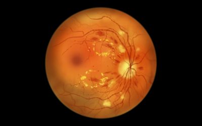
A dilated eye exam by an ophthalmologist serves as the gold standard for detecting diabetic retinopathy. During this painless procedure, your eye doctor places special drops in your eyes to widen your pupils. This provides a clear window into your retina’s blood vessels and other critical structures.
Once your pupils dilate, your doctor examines your retina for microaneurysms, hemorrhages, and abnormal blood vessel growth. This examination can reveal signs of the condition. For example, microscopic bleeding appears as red spots, lipid leakage shows up as yellow deposits, and cotton wool spots appear as white feathery lesions that indicate oxygen-starved retina.
Your doctor might recommend additional tests for more detailed assessment:
- Optical Coherence Tomography (OCT) creates cross-sectional images of your retina to detect fluid buildup
- Fluorescein Angiography uses special dye to highlight damaged blood vessels and areas of poor circulation
Latest treatments: injections, laser, and surgery
Treatment approaches have advanced significantly in the past decade, with options tailored to your specific stage of retinopathy.
Anti-VEGF injections remain the primary treatment for center-involved diabetic macular edema. Medications like faricimab-svoa (VABYSMO™) and aflibercept (EYLEA®) show excellent results. SUSVIMO™, FDA-approved in 2025, delivers continuous medication through a refillable eye implant requiring only one refill every nine months.
Laser treatments continue playing a crucial role in protecting your vision. A focal laser treats specific leaking vessels while panretinal photocoagulation (scatter laser) shrinks abnormal blood vessels to prevent bleeding and further damage.
For advanced cases, vitrectomy surgery removes blood and scar tissue from inside your eye when laser treatment isn’t sufficient.
The critical role of early detection and follow-up care
Regular eye exams and screenings help protect your vision before symptoms appear. The American Diabetes Association recommends eye exams within five years of diagnosis for type 1 diabetes and immediately following diagnosis for type 2 diabetes.
Don’t wait for vision changes to schedule your eye exam. Early detection and treatment can reduce blindness risk by 95%. Follow-up visits, typically every 2-4 months depending on your condition’s severity, allow your doctor to monitor progression and adjust treatment accordingly.
Schedule a comprehensive eye exam at Chang Eye Group in Pittsburgh, especially if you notice any vision changes. Your prompt action today determines how well you’ll see tomorrow.
Taking Action to Protect Your Vision
Your vision deserves protection through every stage of diabetes. The warning signs that we have outlined in this article, from blurry vision to sudden sight loss, demand immediate attention rather than dismissal as minor inconveniences. Recognizing these symptoms early could make the difference between preserving your sight and facing permanent vision loss.
More than 90% of diabetes-related vision loss can be prevented with prompt intervention. Yet many people with diabetes delay seeking care, putting their eyesight at unnecessary risk. Your prompt action when symptoms appear could save your sight.
Don’t wait for multiple symptoms to appear before taking action.
Blurry vision, floaters, difficulty seeing at night, or changes in color perception aren’t just inconveniences. These are messages from your eyes asking for help. Don’t wait for multiple symptoms to appear before taking action.
Regular dilated eye exams remain your strongest defense against diabetic retinopathy. Schedule these appointments according to your doctor’s recommendations, typically annually or more frequently based on your risk factors. Maintaining tight control of your blood sugar, blood pressure, and cholesterol provides additional protection for your retinal blood vessels.
Your eyesight is irreplaceable. The steps you take today to monitor and protect your vision will benefit you for years to come. Schedule an eye exam with Chang Eye Group at our convenient locations in Pittsburgh if you’ve noticed any vision changes or need your regular diabetic eye screening. Your vision health deserves expert care, and early detection makes all the difference in protecting your sight for the future.